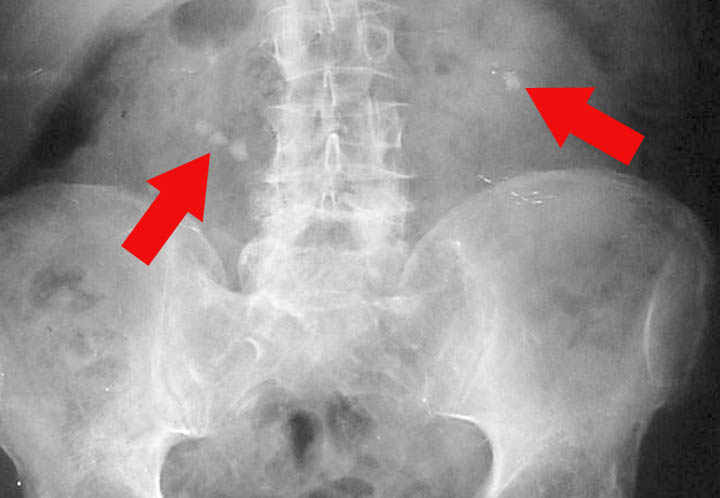
Stones can really be in the wrong place. But today we are not concerned with stumbling blocks, but with stones deep in our body. Often they do not cause us any complaints and are only discovered by chance. But they can also cause painful colics. We are talking about kidney stones. Two to five percent of the total population collects such stones.

Formation
Kidney stones develop when substances accumulate in the urine that are capable of stone formation. The most common are calcium oxalate stones, followed by uric acid stones. The struvite stones are composed of magnesium ammonium phosphate. The rare cystine stones are formed at elevated excretion of the amino acid cystine in the urine.
Increased concentrations of such urine components can result from a lack of fluids, especially in hot areas and in case of long-lasting diarrhea. Diet also plays a role. Eating plenty of dairy products causes an excess of calcium in the urine. If large quantities of meat and sausage is eaten, a lot of purines are produced which are broken down into uric acid. This is precipitated if the fluid intake is too low and, in addition to gout, it can lead to uric acid stones. Drinking plenty of green and black tea increases the absorption of oxalic acid, and calcium oxalate stones will be formed in the presence of calcium.
Lack of exercise in the case of prolonged bed rest or old age can lead to an increased breakdown of calcium from the bones, which in turn promotes stone formation.
Certain metabolic diseases promote stone formation. If the parathyroid gland is overactive (hyperparathyroidism), too much calcium is excreted.
In the case of primary hyperoxaluria, a congenital enzyme disorder, more oxalic acid is found in the urine. This can lead to stone formation.
Diagnosis
A clinical distinction is made between kidney, ureter and bladder stones. If the stones remain in place in the kidney, the patients are usually symptom-free and the stones are discovered only by chance during an X-ray or ultrasound examination. But if they migrate into the ureter, a renal colic occurs. The pain comes like a bolt of lightning out of the blue, with strong labor-like pains, radiating in the lower abdomen, the groin and the genitals. It is accompanied by nausea and vomiting. The patients are very restless and throw themselves around or wander up and down. Large renal pelvis limb stones that cannot be excreted do not cause colics, but only unspecific pain that is interpreted as lumbago. Irritation of the mucous membrane can lead to small amounts of blood in the urine.
Ultrasound and X-ray examinations are used for diagnosis. Stones show up as white reflections with a shadow. At the same time, an elimination urogram is carried out. A radiopaque contrast agent makes the entire urinary system visible, shows the extent of a urinary obstruction and provides information about the type of stones.

Laboratory exams are also used to differentiate the diagnosis. Uric acid, electrolytes (especially calcium) and creatinine are measured in the blood and a hemogram is performed. The pH value of urine is measured. In acidic urine (pH 5) uric acid or urate crystals precipitate, in alkaline (pH 7) phosphate crystals. If the stones injure the mucous membrane, blood is found in the urine.
Complications
When the urinary tract is blocked, bacteria can migrate into the urinary tract. The urine is an ideal breeding ground for bacteria. If the kidneys are infected, urination problems, fever and chills occur. If the bacteria enter the bloodstream, this can lead to blood poisoning. Urinary stasis can cause the renal pelvis to expand.
Therapy
In the case of stones less than 5 mm in size with a smooth surface and no signs of infection, spontaneous elimination can be awaited under the control of ultrasound, urine sediment and hemogram. You should drink plenty of water and exercise a lot, especially climbing stairs and hopping.
Renal pelvic stones are now treated with ESWL (extracorporeal shock wave lithotripsy). The urinary stone is located with X-rays or ultrasound in order to bring it into the focal point of the shock waves. The waves are generated outside the body and the radiation bundled onto the stone. It is smashed by pressure and tension waves. The intensity and number of strokes of the wave are precisely matched to the size and hardness of the stones. This creates stones or sand that can be eliminated spontaneously. The disposal must be controlled. Urine is collected and examined. It is not always possible to smash the stones in one sitting. Sometimes ESWL needs to be repeated or resorted to other treatments.
MIL (minimally invasive percutaneous nephrolitholapaxy) is often the therapy of choice. A small incision is made in the skin under anesthesia, a puncture canal is widened and an endoscope is inserted into the kidney. Kidney stones and smaller fragments can be removed with grasping forceps.
Open surgical interventions for kidney or bladder stones are rarely performed today. For example, if the procedures listed above are unsuccessful or if other complications occur, such as bleeding or injuries to neighboring organs.
Drug dissolution of the stones is used for cystine and uric acid stones. With allopurinol the uric acid level can be lowered.
Follow-up Care for Kidney Stone Patients
Around every fifth patient treated for kidney or bladder stones will have a relapse. That is why the urine must be sieved during treatment and the excreted particles analyzed, so that the patient can be advised precisely according to the type of his stones. Adequate hydration is very important for all kidney stone patients. In addition, special measures apply to the different types of stones.
- With calcium oxalate stones, the consumption of dairy products, cheese, chocolate, spinach, as well as black and green tea should be restricted. Orange juice helps to reduce stone formation.
- In the case of calcium phosphate stones, the consumption of milk, cheese and citrus fruits must be restricted. Currant juice is the preferred drink.
- In the case of uric acid stones, it is important to ensure that the urine is alkalized. This can be achieved by taking K citrate. The patient adjusts the pH of the urine himself with test strips to 6.2 to 6.8. The diet should be low in purine, i.e. contain little meat, sausage and legumes.
- For cystine stones, the urine needs to be adjusted to around 7.5 to 7.8 pH. With a good supply of vitamin C, the cystine can be converted into the more easily soluble cysteine, thus preventing stone formation.
Prevention

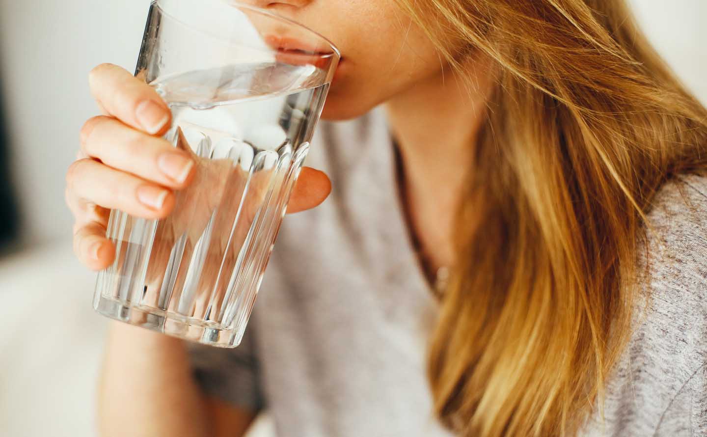
The best prevention is drinking plenty of liquids, and pure water is best suited for this. For a change, you can drink either orange or currant juice, depending on the type of stone. One should rather avoid mineral water. They could be high in calcium or even acidic. The fluid intake must be regularly distributed throughout the day so that the urine is diluted. This way we avoid oversaturation with stone-forming substances.

Stay Always Up to Date
Sign up to our newsletter and stay always informed with news and tips around your health.

Esther Neumann studied Nutrition at the University of Vienna. Since then she served as an author for the health magazine “Leben und Gesundheit” and conducted health lectures in various locations of Austria.
Leave a Reply